40b. That lead is a strongly marked predisposing cause of gout there can be no doubt. Garrod is not at present prepared to assert that the metal cannot of itself occasionally lead to the development of gout. Charcot, with a full knowledge of the views of English writer, has come to the conclusion, founded on his experience of lead – poisoning in France, that though it is a predisposing cause of gout, he has not med with any proof that independent of factor (e. g. excess of meat and beer) it has the special power to create gout. Renaut state that since then Charcot noticed a case under Pidoux’s care in which they were unable to find any cause whatever for the gout except the action of lead. G. Moore, who has had a large experience in lead diseases in the Staffordshire Potteries, writes: “My conviction is that when a man the victim of plumbic impregnation is, as he may be, seized with gout or with rheumatism, the event is not a sequence” (Brit. Journ. of Hom., xxiv, 76). As far as present evidence goes it is more probable that lead predisposes to but does not create gout. Lead arthralgia has no resemblance to gouty swellings and pain. (Ibid.)
41a. The pains of lead colic affect the uterus and vagina, they have been compared to severe labour pains; during the continuance of the pains the vagina is much contracted. Dr. W. Neftel, of New York, has published a brief outline of vaginismus occurring in three young American ladies, of good position, who had for long employed a cosmetic heavily charged with lead. One lady had been married a year, but coitus could not be effected; the two other cases were unmarried. But for his experience in the first case he would have viewed it as an hysterical hyperaesthesia. The only sign of lead – poisoning he reports is paralysis of the extensors of the hands; no mention is made of colic. As the paralysis disappeared, so in proportion did the vaginismus. Treatment: galvanism and iod. pot (Centralbl. f. die med. Wissensch., 1868, p. 819).
41b. The influence of lead on menstruation is not sufficient investigated. Young girls working in white – lead manufactories, long before they get colic, suffer from retarded menstruation, and in older subjects there is suppression of the menses. All show marked anaemia – chlorosis, and are styled by their neighbours “white – lead ghosts.” Hysterical states are extremely prevalent, and the paroxysms particularly violent; chorea and epileptiform convulsions are alarmingly prevalent (LEWIS, Medorrhinum Times and Gaz., 1872, ii, 538). Dowse, from a large experience among women working in the white – lead works in the north – east of London, states that amenorrhoea is very frequently and rapidly produced in the married and unmarried (Ibid., 1876, i, 357).
41c. According to Constantin Paul (Mem. de la Soc. de Biologie, 3 Serie, t. iii.), women who work with lead, especially polishers, are subject to metrorrhagia; this, however, is attendant on abortion. His investigations in this direction shows the unfortunate results of lead on the pregnant uterus and offspring. Of 141 pregnant women whose husbands worked in lead, 82 aborted, 4 had premature delivery, and 5 had stillborn children. Of the 40 living children 20 died within a year, 15 between one and three years. Out of 43 pregnancies in women who had suffered from lead – poisoning there were 32 premature births, 3 stillborn children, and 2 exceedingly pale. Out of 27 pregnancies occurring in five women, 22 aborted – 4 children died – 1 living. One woman, who had aborted five times, left off working in lead, and afterwards bore a fine healthy child. The influence of the father seems less marked than that of the mother. Roque, in a series of observations carried out at the Salpetriere and Bicetre, traced a number of cases of idiocy and epilepsy among the children of parents who had suffered from lead – poisoning, and were temperature. He traced the history of sixteen families in which one or more of the children were affected as above. When the parents had ceased to work in lead, and recovered their health, the children born thereafter were healthy and free from brain disease. (Ibid.)
42. Morbid anatomy. – a Post – mortem appearances in the digestive organs after chronic lead – poisoning are not characteristic. According to Kussmaul and Mayer the gastro – intestinal membrane gives evidence of chronic catarrh. The gastric glands are sometimes atrophied or the seat of fatty degeneration (?). The same lesion with atrophy of the mucous membrane extends to the small intestines and upper part of colon. Peyer’s glands are affected by fatty degeneration. The cellular coats of the stomach and intestines are much thickened. The muscular fibres of the intestines show fatty degeneration in a number of points; more or less limited or wide – spread contraction of the intestines. Tanquerel and Segond drew attention to the condition of the sympathetic ganglia. Kussmaul and Mayer confirm this by describing a proliferation and sclerosis of the connective tissue of several of the sympathetic ganglia, principally in the solar plexus.
42b. The knowledge on the subject of the alterations existing in lead palsy imperfect. Such authors as Charcot, Gombault, Westphal, Vulpian, and Raymond, though in accord as to the condition of the muscular tissue, have different views as to the nervous lesions. Kussmaul and Mayer first noticed a peculiar rigidity of the muscles. This has been confirmed by the observations of Gombault, who describes three different conditions in the muscles of those dying from lead paralysis and cachexia: – Ist, muscles pliant and healthy; 2nd, of yellow colour and atrophied; 3rd, some like smoked flesh, so hard and rigid that a long muscle, detached from its insertions, could be held horizontally without bending. True fatty degeneration has very rarely been seen. Ollivier and Lancereaux have observed granular degeneration, consisting partly in simple atrophy, the muscle preserving its structure but showing diminution of contractile substance. The adipose vesicles increase between the primitive muscular fasciculi, which then become yellow and pale. Side by side with these changes, and succeeding them, the nuclei on the internal face of the sarcolemma are multiplied, pushing back the contractile substance, forming accumulations at various points, giving the muscular fibre a mossiliform appearance. The examination of the central nervous system has given no results. Raymond and Vulpian found in one case that the cervical portion of the spinal cord was altered. The anterior roots, as also the muscular nerves, were normal; the anterior born of the cord presented on its external aspect various atrophied cellules, shrunken, deprived of nuclei, and everywhere pierced with small vacuoles. Lancereaux has pointed out a granular adipose degeneration of the myeline, and the spinal cord showed a little atrophy in the centre of the anterior roots; there was no lesion of the horns of the grey substance. Gombault has been unable to find any changes in the medulla. Dejerene (Lond. Medorrhinum Rec., 1879, p. 112), from a careful examination of five cases, confirms the description above given of the intra – muscular nerves and radial nerve. In two cases he found alterations in the anterior cord. In two cases he was able to trace distinctly the degeneration of a certain number of the nervous tubuli similar to that met with in the intra – muscular nerves. In one of these cases, where the patient was only paralysed on one side, the roots had degenerated on the corresponding side, the other being perfectly healthy. In the three other cases no distinct change could be found. [ “In speaking of the pathogenesis of lead paralysis I have sought to show that its origin is probably spinal instead of peripheral, and E. Remark has established this more accurately and in a more detailed manner by the sifting and critical examination of a larger amount of material. He comes to the conclusion that quite circumscribed alterations in the grey anterior horns probably lie at the foundation of lead paralysis. These alterations might well be of a degenerative or chronic inflammatory nature, but are, as a rule, capable of resolution. Bernhardt has also recently given in his adherence to this view.” (ERB, in Ziemssen’s Cyclopoedia, Xiii, 715; see also xi, 546.) ] (Ibid.).
Experiments on animals
1. ORFILA found that in large doses the acetate acted on dogs as an irritant, and caused vomiting, pain, and death. When the action was slower, and absorption took place, an affection of the nervous system was observed, marked by difficult progression, and, in some cases, convulsive movements. When injected into the jugular vein 13 gr. killed a dog almost immediately, death being preceded by no other symptom except convulsive respiration. Another was killed by 5 gr. in as many day; the leading symptoms were weariness, languor, staggering, and slight convulsions, none of which symptoms appeared till the 3rd d. In neither animal could be find any morbid appearance on dissection. (Opium cit.)
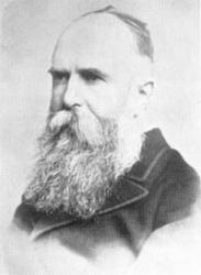
Hughes was a great writer and a scholar. He actively cooperated with Dr. T.F. Allen to compile his 'Encyclopedia' and rendered immeasurable aid to Dr. Dudgeon in translating Hahnemann's 'Materia Medica Pura' into English. In 1889 he was appointed an Editor of the 'British Homoeopathic Journal' and continued in that capacity until his demise. In 1876, Dr. Hughes was appointed as the Permanent Secretary of the Organization of the International Congress of Homoeopathy Physicians in Philadelphia. He also presided over the International Congress in London.